
Diagnostics of the Tularemia
Differential diagnosis, Tularemia should be distinguished from coccal, tuberculous and other etiology lymphadenitis, lymphogranulomatosis, pneumonia (in pulmonary form), lymphosarcoma, felinosis, infectious mononucleosis, ornithosis, Q fever, in natural foci – from plague.
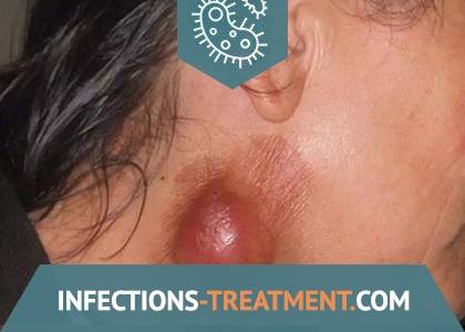
Tularemia lymphadenitis is distinguished by the subsiding of pain with an increase in bubo, weak or absent phenomena of periadenitis, slow resorption or sclerosis, and with the suppuration of bubo, the creamy character of pus. Of the signs of the disease common to all forms of tularemia, attention is drawn to high prolonged fever, relative bradycardia, hepatolienal syndrome, and the possibility of exanthema of a different nature.
With the ulcerative-bubonic form, the development of the primary affect at the site of the introduction of the pathogen in the form of successively replacing spots, papules, vesicles, pustules, ulcers is characteristic. With the ocular-bubonic form of tularemia, the mucous membranes of the eyes are affected in the form of conjunctivitis, papular, and then erosive-ulcerative formations with the separation of yellowish pus. Angina in the angina-bubonic form of the disease is often distinguished by a one-sided character, moderate pain in the throat, adhesion of the tonsils with the surrounding tissue, hard-to-remove grayish-white plaques on their surface, and in the future – the formation of deep ulcers that slowly heal with scarring. Lesions of the mesenteric lymph nodes in the abdominal form are clinically manifested by severe abdominal pain, nausea, occasionally vomiting, anorexia. The bronchitic variant of the pulmonary form of tularemia is distinguished by the defeat of the bronchial, mediastinal, paratracheal lymph nodes, tularemia pneumonia – a rather severe acyclic course, a tendency to develop complications (bronchiectasis, abscesses, pleurisy, cavities, gangrene of the lungs).
Laboratory diagnostics
In the early days of the disease, moderate leukocytosis, neutrophilic shift to the left, and increased ESR are noted in the peripheral blood. In the future, leukocytosis can replace leukopenia with lymphocytosis and monocytosis. In clinical practice, serological research methods are widely used – RA (minimum diagnostic titer 1: 100) and RNGA with an increase in antibody titer in the dynamics of the disease. ELISA on a solid-phase carrier is positive from 6-10 days after the disease, the diagnostic titer is 1: 400; in sensitivity, it is 10-20 times higher than other methods of serological diagnosis of tularemia. Also, the setting of a skin-allergic test with tularin is common: 0.1 ml of the drug is injected intradermally into the middle third of the forearm from the inside; the result of the reaction is taken into account after 1-2 days. The test is highly specific and effective already in the early stages (on the 3-5th day) of the disease. Its positive result is expressed in the appearance of infiltration, soreness and hyperemia with a diameter of at least 0.5 cm. It should be borne in mind that the test can also be positive in persons who have had tularemia.
Bacteriological diagnosis of tularemia is of secondary importance, since isolation of the pathogen from blood or other pathological materials is difficult and not always effective. Isolation of the pathogen is possible in the first 7-10 days of the disease, but this requires special environments and laboratory animals. Isolation of the pathogen, as well as the setting of a biological test with infection of white mice or guinea pigs with punctate of buboes, blood of patients, discharge of the conjunctiva and ulcers are possible only in special laboratories for working with pathogens of especially dangerous infections. Molecular genetic method: PCR is positive in the initial febrile period of the disease and is a valuable method for early diagnosis of tularemia.
Treatment of Tularemia
Etiotropic therapy involves the combined use of streptomycin 1 g / day and gentamicin 80 mg 3 times a day intramuscularly. You can prescribe doxycycline 0.2 g / day by mouth, kanamycin 0.5 g 4 times a day and sisomycin 0.1 g 3 times a day intramuscularly. The course of antibiotic treatment is continued until the 5-7th day of normal body temperature. The second line of antibiotics includes III generation cephalosporins, rifampicin and chloramphenicol.
They carry out detoxification therapy, antihistamines and anti-inflammatory drugs (salicylates), vitamins, cardiovascular agents are shown. For local treatment of buboes and skin ulcers, ointment dressings, compresses, laser irradiation, and diathermy are used. When bubo suppurates, it is opened and drained.
Patients are discharged from the hospital after clinical recovery. Long-term non-absorbable and sclerosed buboes are not a contraindication for discharge.
Prevention of Tularemia
Epizootological and epidemiological surveillance
Includes constant monitoring of the incidence of humans and animals in natural foci of tularemia, circulation of the pathogen among animals and blood-sucking arthropods, monitoring the state of immunity in humans. Its results form the basis for planning and implementing a complex of preventive and anti-epidemic measures. Epidemiological surveillance provides for an epizootological and epidemiological examination of natural foci of tularemia, generalization and analysis of the data obtained in this case, causing epidemic manifestations in natural foci of tularemia in the form of sporadic, group and outbreak of human morbidity.
Preventive actions
The basis for the prevention of tularemia is made up of measures to neutralize the sources of the causative agent of the infection, neutralize the factors of transmission and carriers of the pathogen, as well as vaccination of threatened contingents of the population. Elimination of the conditions for human infection (general sanitary and hygienic measures, including sanitary and educational work) has its own characteristics for various types of morbidity. In case of transmissible infections through bloodsucking, repellents, protective clothing are used, and the access of the unvaccinated population to disadvantaged areas is limited. The fight against rodents and arthropods (deratization and disinsection measures) is of great importance. For the prevention of alimentary contamination, swimming in open water should be avoided, and only boiled water should be used for household and drinking purposes. When hunting, you need to disinfect your hands after skinning and gutting hares, muskrats, moles and water rats. Vaccination is carried out in a planned manner (among the population living in natural foci of tularemia, and contingents at risk of infection) and according to epidemiological indications (unscheduled) when the epidemiological and epizootic situation deteriorates and the threat of infection of certain population groups arises. For immunoprophylaxis, a live attenuated vaccine is used. Vaccination provides the formation of stable and long-term immunity in vaccinated (5-7 years and more). Revaccination is carried out after 5 years for the contingents subject to routine vaccination.
Activities in the epidemic focus
Each case of human disease with tularemia requires a detailed epizo-otologic-epidemiological examination of the focus with the elucidation of the route of infection. The question of hospitalization of a patient with tularemia, the timing of discharge from the hospital is decided by the attending physician purely individually. Patients with abdominal, pulmonary, ocular-bubonic and anginal-bubonic, as well as moderate or severe cases of ulcerative-bubonic and bubonic forms should be hospitalized for clinical indications. Patients are discharged from the hospital after clinical recovery. Long-term non-absorbable and sclerosed buboes are not a contraindication for discharge. Dispensary observation of a patient who has been ill is carried out for 6-12 months in the presence of residual effects. Separation of other persons in the outbreak is not carried out. As a measure of emergency prophylaxis, antibiotic prophylaxis can be carried out with the appointment of rifampicin 0.3 g 2 times a day, doxycycline 0.2 g 1 time per day, tetracycline 0.5 g 3 times a day. Disinfection is carried out in the patient’s home. Only things contaminated with patient secretions are subject to disinfection.