
What is Echinococcosis?
Echinococcosis is a rare chronically parasitic disease that occurs as a result of exposure to the body of the larval form of the tape helminth Echinococcus granulosus.
Echinococcosis is widespread throughout the world. According to statistics, the population and animals of the southern countries are most intensely affected: countries of South America (Uruguay, Paraguay, Argentina, Chile, Brazil), Australia and New Zealand, North Africa (Tunisia, Algeria, Morocco, Egypt), Southern Europe (Italy, Greece, Cyprus, Turkey, Spain, Yugoslavia. Bulgaria, France), then – the southern part of the USA, Japan, India, the former USSR. As you move from south to north, the prevalence decreases. In the territory of the former Union, echinococcosis is widespread in those republics and regions where animal husbandry is developed, mainly sheep breeding – in the North Caucasus, Transcaucasia, Kazakhstan, Kyrgyzstan, Uzbekistan, Moldova (the incidence of the population is 1.37 – 3.85 per 100,000), in Russia – Bashkortostan, Tatarstan, Stavropol, Krasnodar, Altai, Krasnoyarsk, Khabarovsk Territories, Volgograd, Samara, Rostov, Orenburg, Chelyabinsk, Tomsk, Omsk, Kamchatka, Magadan, Amur Regions and Chukotka Autonomous Okrug.
In Ukraine, echinococcosis is more often recorded in the southern regions – Odessa, Crimean, Kherson, Nikolaev, Donetsk, Zaporizhzhya, in the rest – sporadic cases.
On the territory of Ukraine, 2 types of foci are recorded: in the southern steppe zone, a “sheep” strain circulates, in the Polesie and forest-steppe, mainly “pig” strain. The defeat of sheep in the Odessa region was 32%, cattle – 20%, pigs – 9%.
Causes of Echinococcosis
The causative agent of human echinococcosis is the larval stage of the echinococcus tapeworm – Echinococcus granulosus.
The sexually mature form of echinococcus is a small cestode 2.5 – 5.4 mm long and 0.25 – 0.8 mm wide. It consists of a pear-shaped scolex, neck and 3-4 segments.
Skoleks is equipped with four suction cups and a crown from two rows of hooks (28 – 50).
Behind the scolex there is a short neck and joints, the first two are immature, the third hermaphroditic and the fourth mature. The mature segment (length 1.27 – 3.17 mm) is filled with a stretched uterus, which is a wide longitudinal trunk with lateral protrusions. The uterus is packed with eggs (400 – 600 pieces), which do not differ in structure from the eggs of bovine and pork tapeworms (teniids), which contain a six-hooked oncosphere inside.
The sexually mature form – echinococcus tapeworm – parasitizes only in animals: dogs, wolves, jackals, foxes, which are the ultimate hosts. The larval stage – an echinococcal cyst – parasitizes in intermediate hosts – various herbivorous and omnivorous ungulates (sheep, goats, cattle, pigs, horses, etc.) and humans.
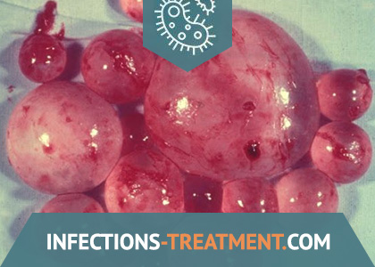
Echinococcal cyst is a bubble of a very complex structure. Outside, it is surrounded by a layered membrane (cuticle), the thickness of which sometimes reaches 5 mm. Under the multilayer cuticular membrane lies a thin inner germinal (germinative) membrane, which produces brood capsules with scolexes, daughter blisters, and also gives rise to the layered membrane.
Brood capsules are small vesicle-shaped formations scattered on the germinal membrane and connected to it by a thin leg. They have the same structure as the main bladder, but with the opposite arrangement of the shells (germinate on the outside, layered on the inside). Each brood capsule contains scolexes attached to its wall, screwed inward and having a typical structure for chains. The bubble is filled with a liquid that acts as a protective nutrient medium for brood capsules and scolexes.
The liquid may contain freely weighed, detached scolexes and brood capsules, the so-called hydatidose sand.
The bladder is gradually covered with a connective tissue membrane. Often in such a maternal cyst, in addition to the above listed elements, there are also so-called daughter bubbles, which have the same structure, and granddaughter bubbles inside them.
Such cysts are observed in humans. Sometimes daughter bubbles do not form inside the maternal cyst, but on the outside. Such bubbles are called exogenous.
Echinococcal cysts formed in animals, as a rule, do not contain brood capsules and scolexes, they are called acephalocysts. In humans, this form does not occur.
In the sheep-breeding areas of the southern zone, the echinococcus circulation follows the pattern: sheep – ›guard dogs accompanying the flock -› sheep.
In the western regions of intensive pig breeding, the echinococcus circulation follows the pattern: pigs – ›dogs -› pigs. The lack of active motor function in the members of the “swine” strain reduces the contamination of dog hair and soil, thereby limiting the conditions of infection of people and animals.
The intensity of invasion transmission is determined, first of all, by the number of sources of invasion of the final hosts and the amount of invasive material released by them – oncospheres and segments.
The oncospheres tolerate temperatures from -30 ° C to + 38 ° C, they remain viable for a month on the surface of the soil in the shade at a temperature of 10 – 26 ° C, but die in the sun at a temperature of 18 – 50 ° C after 1-5 days. In the grass at a temperature of 14 – 28 ° C, they die no earlier than after 1.5 months. Oncospheres tolerate low temperatures, at which they can persist for several years, but are very unstable to dry.
The circulation of invasion in echinococcosis is carried out according to the well-known pattern: the source of invasion (the final hosts are carnivores) – ›the external environment polluted by the oncospheres and segments of the parasite, -› the intermediate host (herbivores, omnivores, infected by larvae) – ›the uninfected final host.
Man – an intermediate host – is a biological dead end.
With human echinococcosis, the dog occupies the main position as the final host. Dogs become infected when they eat meat from slaughterhouses, slaughter areas, kitchens, when they feed them confiscated slaughterhouses or organs of animals slaughtered at home affected by larvocists. It is also possible that dogs are infected by feeding them hunting products – affected organs or corpses of wild herbivores.
Ways of infection of intermediate hosts are also different, herbivorous farm animals become infected by swallowing eggs, segments of helminth with grass, hay, water, contaminated with feces of invaded dogs. Pigs, being coprophagous, become infected by eating dog feces. The main role in infecting a person through dirty hands is played by communicating with invasive dogs, the hair and tongue of which can contain eggs and segments of echinococcus chains. Healthy animals can also transmit invasion to humans as mechanical carriers of eggs that contaminate their hair, tongue when licking an infected dog.
Human infection is not excluded when eating unwashed vegetables, berries, fruits contaminated with feces of dogs containing oncospheres.
A person can also become infected from wild carnivores during hunting when cutting hides, making fur clothes, as well as eating unwashed wild berries and drinking water from natural reservoirs.
In sheep-breeding areas, where the pathogen is mainly circulated between dogs and sheep, risk groups include shepherds, shepherds accompanying flocks, and sheared sheep’s wool and family members.
Pathogenesis during Echinococcosis
Echinococcosis develops in connection with the introduction and growth in various organs of the tape worm larva – echinococcus.
A person is infected with echinococcosis mainly orally, and due to the hematogenous pathway of the oncosphere, it can affect any organ, any tissue, but most often the liver (44 – 85%), then the lungs (15 – 20%) in more rare cases, in a large circle of blood circulation – kidneys, bones, brain and spinal cord and other organs.
In the affected organs, one cyst may develop, or how many – multiple echinococcosis, depending on the introduced oncospheres.
The pathological effect of echinococcus is due to the mechanical and sensitizing effect of the growing larva. The sizes of cysts are from 1 – 5 cm in diameter to giant cysts containing several liters of fluid. The mechanical effect of such a cyst leads to impaired function of the affected organ. Localization and size determine the main symptomatology and severity of the disease.
Sensitization of the body by parasite metabolism leads to the development of immediate and delayed hypersensitivity. A striking manifestation of an allergic reaction of an immediate type is eosinophilia and urticaria as a result of leakage of echinococcal fluid, and in more severe cases (when opening the bladder) anaphylactic shock. In the late stages of the disease, especially with multiple echinococcosis, an important role is played by immunopathological reactions.
The primary owner of echinococcosis are dogs, wolves, jackals, foxes, etc. The parasite that lives in their small intestine consists of a head, neck and segments – the posterior and largest, the fourth, is mature. Such a mature segment, separated from the worm, throws eggs that are excreted with feces. More often than not, a dog, being a carrier of helminth (worm), serves as a source of contamination of echinococcus eggs in pastures, ponds, premises for animals and human dwellings.
From the moment a joint or its eggs (the so-called oncosphere) is swallowed, the period of development of the parasite larva begins. Digestive juices help the embryo to free itself from the membranes and with the help of its hooks penetrates the mucous membrane of the gastrointestinal tract. Further, with the flow of blood or lymph, it spreads to the organs (liver, lungs, kidneys, muscles) and, already sowing in their tissues, turns into a larva. By the end of 2 weeks, it takes on a bubble structure.
After 5 months. the resulting bubble has a diameter of 5 mm. In the future, the vesicle grows slowly over the years, and gradually, after 20-25 years, reaches a large size, with a capacity of 10 l or more: a connective tissue capsule with chitin walls. The cavity of this cyst is filled with a slightly yellowish neutral reaction liquid containing sodium chloride, grape sugar, tyrosine, succinic acid, albumin, etc. The chitinous membrane consists of two layers: the outer dense (cuticular) thickness up to 0.5 cm and the inner (germinative) germinal from which are formed in large quantities, sometimes up to 1000, daughter bubbles.
Pathological changes in the human body are associated with mechanical pressure on the organs of a growing cyst. The vital products of the parasite irritate the surrounding tissues, causing their chronic inflammation.
Symptoms of Echinococcosis
There are four stages of echinococcosis:
– the first is latent (initial asymptomatic), from the moment of invasion of the oncosphere (penetration into the body) until the appearance of subjective signs;
– the second – mild, mainly subjective disorders;
– third – pronounced objective symptoms and
– fourth – complications.
The duration of the stages, given the slow growth of an echinococcal cyst, is difficult to establish. It can only be noted that the rate of increase in symptoms is associated with the localization of echinococcus. So, for example, a cyst developing in the peripheral parts of the liver parenchyma for many years may not give any sensations, but if it develops near the gate of the liver, then, squeezing the liver passages, quickly causes obstructive jaundice, and squeezing the portal vein, leads to the development of ascites .
Clinical manifestations in the initial stages of the disease are usually scarce, they are detected when the cyst reaches a significant size or, due to its location, compresses an important organ and leads to a violation of its function. Echinococcal cyst of internal organs (liver, kidney, spleen, etc.) is usually recognized when a tight-elastic tumor is palpated, and damage to the lungs and bones is determined on x-rays in the form of cystic formations.
Echinococcosis of the liver is more common. With uncomplicated liver echinococcosis, a growing cyst stretches the capsule of the organ, causing dull, aching, less often – paroxysmal pain.
According to the classification of A. V. Melnikov, there are 3 stages of the course of invasion.
The first is the initial asymptomatic – from the moment of infection to the manifestation of the first clinical signs of the disease. The second stage is the appearance of the symptoms of the disease: patients complain of weakness, decreased performance, poor appetite, nausea and vomiting, sometimes stool disturbance. Against this background, there are sensations of pressure and heaviness in the right hypochondrium, epigastrium, and sometimes sharp, severe dull pains. The liver on palpation is enlarged slightly painful, with a superficial location of the cyst – soft, elastic, with calcification – woody density. Squeezing the portal or inferior vena cava, as well as squeezing the intra- and extrahepatic bile ducts leads to the development of obstructive jaundice.
The third stage is the stage of pronounced pathological changes and complications: the development of an abscess, a possible rupture of a cyst with fever with chills, pain in the upper abdomen.
In the hemogram – leukocytosis with a stab shift, eosinophilia is possible.
Cyst rupture – the most serious complication of echinococcosis occurs with bruise, fall, sudden movement, severe coughing. Clinically, perforation of the cyst can manifest itself as a complex of allergic reactions up to the development of allergic shock. Especially dangerous dissemination of echinococcal cysts.
Echinococcosis of the lungs – the second most frequent manifestation of invasion, can simulate any lung disease of another etiology.
The first stage – the stage of an unopened echinococcal cyst – is associated with the growth of the bladder, compression of lung tissue, blood vessels, bronchi, and pleura involvement. Patients are concerned about shortness of breath, hemoptysis, chest pain, severe cough, especially at night, initially dry, then turning wet with mucopurulent sputum. Visually – chest deformity, smoothness of the intercostal spaces. Percussion – dullness of sound, auscultation – weakening of breathing, pleural friction noise, etc.
The second stage is associated with the opening of the cyst. With a breakthrough in the bronchus (more often), a strong cough, suffocation, cyanosis, severe allergic reactions, the development of aspiration pneumonia appear. With a breakthrough in the pleura, the pericardium may cause anaphlactic shock and sudden death.
In cases of echinococcosis of other organs (spleen, kidneys, bones, muscles), symptoms simulating a tumor process predominate.
Important common symptoms of echinococcosis are periodically developing signs of an allergic reaction (urticaria, etc.), which is usually associated with absorption of echinococcal fluid in case of cyst shell tears or as a result of surgery. For echinococcosis, as, incidentally, for other helminthic diseases, eosinophilia is characteristic, reaching 10-25%.
Dangerous complications of an echinococcal cyst are suppuration or rupture of it with contamination of the abdominal, pleural or any other cavity.
Diagnosis of Echinococcosis
Diagnosis of echinococcosis often presents significant difficulties. In recent years, in addition to general clinical research methods, contrast methods, such as transumbilical portohepatography, selective angiography of the celiac artery, scanning with radioactive isotopes, which help to diagnose liver echinococcosis, computed tomography, have become more widely used.
The range of methods used for immunodiagnostics is wide.
A specific laboratory reaction is the Katsoni reaction, which with echinococcus is positive in 89-90% of cases. The technique for producing the Katsoni reaction is as follows: 0.2 ml of sterile echinococcal fluid is administered intradermally. With a positive reaction, redness appears at the injection site, and then continuous intense redness (cutaneous anaphylaxis).
Serological reactions with a specific antigen are used: NRIF (sensitivity and specificity according to the literature 88 and 98.6%, respectively), NRGA (specificity 79.26%, sensitivity 88.68%); Elisa – enzyme immunoassay – (specificity – 78.52% and sensitivity – 90.57%); Sandwich – Elisa for the detection of specific antigens of E. granulosus in feces, immunoblot, etc.
Previously used reactions: scolexoprecipitation, latexagglutination are not used in practice.
Echinococcosis Treatment
Echinococcus extraction is possible only by surgery. There are several methods of operation:
1) radical echinococcectomy, i.e. complete removal of echinococcal cyst together with its fibrous membrane,
2) opening the cyst with the removal of fluid, all daughter bubbles and the chitinous membrane with rubbing the formed cavity with a formalin disinfectant solution and plugging, draining or suturing it tightly.
When opening an echinococcal cyst, special attention is paid to isolating the body cavities and tissues from the echinococcal fluid, since its ingress into the cavities (abdominal, chest, etc.) or on the wound walls can lead to seeding.
Prevention of Echinococcosis
The complex of veterinary and medical measures for echinococcosis is aimed primarily at identifying and eradicating the source of invasion. In accordance with official recommendations, we are talking about reducing the number of guard dogs, their registration, registration and destruction of stray animals.
Veterinary specialists of farms carry out preventive deworming of service dogs from December to April every 45 days, from May to November – every 30 days, the rest – once a quarter. These measures must be carried out with respect to personal dogs. Deworming is carried out at special sites where the separated feces are collected in a metal container and neutralized: (boil for 10 – 15 minutes, pour for 3 hours with a 10% solution of bleach, the soil is treated with a 3% solution of carbathion (4 l per 1 m2 )
To prevent infection of dogs, it is necessary to comply with the rules for slaughtering farm animals and ensure the destruction of affected organs, as well as blocking dogs from entering meat processing plants, slaughterhouses, and cattle burial grounds.
Measures to prevent infection of dogs also include such mandatory recommendations as: raising the veterinary-sanitary level of farms; construction of recycling pits, cattle cemeteries; compliance with the rules of storage and transportation of animal corpses; slaughter of animals only in appropriate places for this, etc.
Medical measures include identification of militant groups invaded by examination (hunters, persons in contact with dogs involved in fur processing, manufacturing of fur products, shepherds) and examination according to clinical indications; deworming and medical observation. Health education is important.
Personal prevention of echinococcosis is to limit contact with dogs, children playing with them, wash their hands thoroughly after contact with animals, before eating after working in the garden, playing in the yard, in the garden, picking mushrooms, do not eat unwashed wild berries Do not drink unboiled water from natural reservoirs.